Glaucoma Clinic
Glaucoma refers to a group eye conditions that affects the pressure within the eye, which damages the optic nerve, the nerve connecting your eye to the brain. The best way to test if you have glaucoma is to have a complete eye examination with your Ophthalmologist. 90% of glaucoma patients in India do not realise that they have the disease. Often called ‘the sneak thief of sight’, people with most forms of glaucoma do not have symptoms until the optic nerve is severely damaged. Early diagnosis and treatment can help in controlling the disease and preventing the permanent loss of vision.
If you are at risk of developing glaucoma you may be required to have periodic eye examinations with multiple tests at regular intervals.The risk factors of developing glaucoma include age >40 years, family history, having an eye power, high eye pressures, diabetes, steroid users, have had an eye injury.
To achieve an accurate assessment, experienced specialists at our glaucoma clinic perform a comprehensive screening that consists of non invasive, pain free procedures. It also serves as a large referral centre for treatment of congenital glaucoma, which is a rare condition in which the eye pressures are high in new borns or in first few months of life.
Our clinic is well equipped with the latest modalities for diagnosis and treatment of the disease to improve the patients quality of life. Treatment is available for all the age groups from the new borns to the elderly

OPEN ANGLE GLAUCOMA
Open-angle glaucoma, the most common form of glaucoma, accounting for at least 90% of all glaucoma cases:
- Is caused by the slow clogging of the drainage canals, resulting in increased eye pressure
- Has a wide and open angle between the iris and cornea
- Develops slowly and is a lifelong condition
- Has symptoms and damage that are not noticed.
“Open-angle” means that the angle where the iris meets the cornea is as wide and open as it should be. Open-angle glaucoma is also called primary or chronic glaucoma. It is the most common type of glaucoma.

ANGLE CLOSURE GLAUCOMA
Angle-closure glaucoma, a less common form of glaucoma:
- Is caused by blocked drainage canals, resulting in a sudden rise in intraocular pressure
- Has a closed or narrow angle between the iris and cornea
- Develops very quickly
- Has symptoms and damage that are usually very noticeable
- Demands immediate medical attention.
It is also called acute glaucoma or narrow-angle glaucoma. Unlike open-angle glaucoma, angle-closure glaucoma is a result of the angle between the iris and cornea closing.
NORMAL TENSION GLAUCOMA (NTG)
Also called low-tension or normal-pressure glaucoma. In NTG the optic nerve is damaged even though the eye pressures are within normal limits.
CONGENITAL GLAUCOMA
This type of glaucoma occurs in babies when there is incorrect or incomplete development of the eye's drainage canals during the prenatal period. This is a rare condition that may be inherited. When uncomplicated, microsurgery can often correct the structural defects. Other cases are treated with medication and surgery.
SECONDARY GLAUCOMA
There are certain other types of glaucoma where there is an identifiable cause for increased eye pressure resulting in optic nerve damage and vision loss. These are called secondary glaucoma. It may be caused by prolonged, indiscriminate use of steroids, severe diabetic retinopathy, retinal vein occlusion, injuries to the eye, inflammation of the eye (uveitis) or advanced cases of cataract.
If you believe you have any of these risk factors get an eye examination done. Always remember to inform your eye doctor about the risk factors that you have. This will help your doctor decide how often you need to get your eyes examined.
Diagnostic Tests for Glaucoma
If You have Glaucoma, See the chart below.

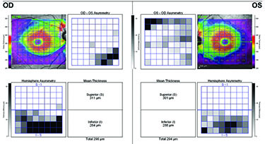
The clinic handles Primary Glaucomas, which are largely inherited, in addition to Secondary Glaucomas caused by trauma, inflammation, Diabetes, Retinal Vascular Disease and hypermature cataracts. Deviations from normal retinal nerve fibre thickness are the earliest structural changes in persons with glaucoma.
Tonometry
Measures the pressure inside your eye
- Non contact tonometer
- Icare tonometer
- Goldmans applanation tonometer
- Perkins tonomter

Gonioscopy
To assess the status of the drainage angle in your eye
- Pachymetry- measure the thickness of your cornea. Thickness can influence your eye pressure and risk of developing glaucoma
- Structural assessment of optic nerve - .

Pachymetry
Measure the thickness of your cornea. Thickness can influence your eye pressure and risk of developing glaucoma

STRUCTURAL ASSESSMENT OF GLAUCOMA
Fundus photography
To document the severity and monitor the changes in the optic nerve head over time

Eidon Fundus Camera

Optical Coherence Tomography (OCT)
Precise measurement of your nerve fibre layer to detect and monitor the changes over time
- Heidelberg Spectralis OCT


Functional Assessment of Optic Nerve
Perimetry - Assessment of field of vision


Electroretinogram - Assessment of early damage

Treatment
The aim of glaucoma treatment is to lower the eye pressures, to preserve the visual function and avoid further damage to optic nerves. It can be achieved by medications or eye drops which may have to be continued for a long term. But sometimes laser or surgical procedures might be required to obtain adequate lowering of the pressures. It is best to discuss all possible options that you may have with your consultant.
Laser procedures available
- Nd YAG laser peripheral iridotomy
- Selective laser trabeculoplasty
- Micropulse laser trabeculoplasty
- Micropulse transcleral cyclo g6 laser therapy
Routinely performed surgical procedures
- Trabeculectomy
- Trabeculectomy + Trabeculotomy.
- Trabeculectomy with anti mitotic agents like Mitomycin-c or 5-FU
- Trabeculectomy with ologen implants,
- Valve /Tube implants
- Combined Trabeculectomy with IOL implantation.
- Bleb needling , bleb repair/ revision
- Non penetrating glaucoma filtering surgeries : deep sclerectomy (including laser assisted)
LASER PERIPHERAL IRIDOTOMY (LPI)
LPI is a procedure used in the treatment of angle closure glaucoma or as a preventive measure in people who are at risk of angle closure glaucoma. Angle closure refers to narrowing of your drainage canal which results in raised eye pressures and optic nerve damage. It is an office based procedure where a tiny opening is created in your iris(coloured part of eye) with the help of laser energy, which help in widening the drainage canal.

SELECTIVE LASER TRABECULOPLASTY (SLT)
SLT is a form of laser procedure which involves application of laser energy to the trabecular meshwork which is the site of aqueous (fluid in the eye) drainage. It helps fluid to drain out of the eye, reducing the eye pressures. It may take 1-3 months for the effect of the treatment. This method can be used as initial treatment in glaucoma or along with eyedrops as an additional IOP lowering measure. SLT has fewer side effects compared to other treatment modalities. It can lower the eye pressures by about 20-30 %. The effect can wear off over several years, at which point the procedure can be repeated with safety


MICROPULSE LASER TRABECULOPLASTY (MLT)
MLT involves application of repetitive low energy laser pulses that are separated by brief rest periods to the trabecular meshwork. This method can be used as initial treatment in glaucoma or along with eyedrops as an additional IOP lowering measure. Post procedure inflammation is less compared to SLT. Treatment risks and damage to the trabecular meshwork is very minimal in this procedure and therefore can be repeated as needed.

MICROPULSE CYCLO G 6 transcleral cyclophotocoagulation
MicroPulse laser therapy is a non incisional procedure with rapid recovery without quality of life impact. It is an effective alternative to expensive medications and invasive surgeries. With MicroPulse, a continuous-wave laser beam is chopped into a train of short, repetitive, low energy pulses separated by a brief rest period which allows the tissue to cool between laser pulses. It has 30-45% reduction in IOP.

TRABECULECTOMY
A surgical procedure, which is effective in lowering the intraocular pressure and prevent further damage of the optic nerve. The procedure involves creating an alternative pathway of drainage of fluid from the eye, which keeps the pressure under control. We may be able to decrease or completely stop you anti-glaucoma medications post surgery.Trabeculectomy procedure will not reverse any loss of vision that has already occurred. It will prevent further loss.
- During the surgery the surgeon may opt to use additional drops or implants to increase the success of the surgery. This includes drops like mitomycin c or 5 flouro uracil (5FU) and ologen implant.
- If you have a significant cataract, the same can be removed in one sitting which means cataract surgery can be combined with glaucoma surgery or we can do it separately.
In case of congenital glaucoma and paediatric glaucoma the surgery is trabeculectomy with trabeculotomy

DRAINAGE IMPLANTS OR TUBE SURGERY
When your eye pressures are not controlled with medications, laser or trabeculectomy surgery, you may have to go on with drainage implants which bypasses your drainage channels. The device consists of a tube and a plate. The tube is inserted into the front chamber of your eye, which drains the fluid into the plate, which is placed under the conjunctiva. The fluid is absorbed by the natural drainage around the plate. The implant is not visible externally, unless you lift the eyelid.

RESEARCH (ONGOING STUDIES)
-
The effect of trabeculectomy on diurnal variation of IOP and water drinking test
-
The effect of central corneal thickness on measurement of intraocular pressure with icare tonometry
-
Retrospective study on Intraocualar pressure drop after phacoemulsification
-
The accuracy of Spectralis OCT to detect bruch’s membrane opening in myopic patients with peri papillary atrophy
-
Awareness and knowledge about the medications among glaucoma patients
-
Outcome of micropulse trans scleral cyclophotocoagulation with MP3 probe Cyclo G6