Clinical Value
Clinical Value
-Assessment of neuroaxonal injury and detection of minute changes over time
High clinical relevance correlating with established clinical criteria
-Associated with physical disability (EDSS) and quality-of-life scores
-Correlated with brain MRI lesions (T1 and T2) and atrophy (BPF)
-Related to disease activity, duration and cognitive decline
-RNFL and ganglion cell thinning can be detected in MS patients with and without optic neuritis
-Excellent reproducibility and sensitivity
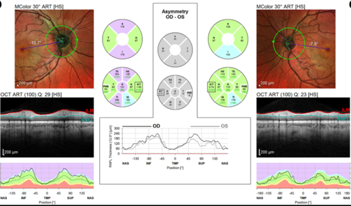
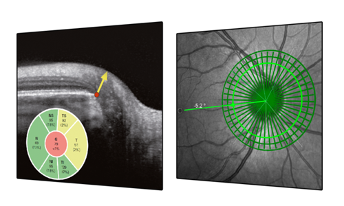
FoBMOC: Automatic Fovea-to-BMO-Center Axis Alignment
Studies have shown that RNFL thinning in MS patients with a history of acute optic neuritis is more likely to be found in the temporal quadrant. The papillomacular bundle (PMB) may be most sensitive to this type of axonal damage. FoBMOC: Automated Fovea-to-BMO-Center Axis Alignment correctly orients the anatomy for PMB measurement accuracy.
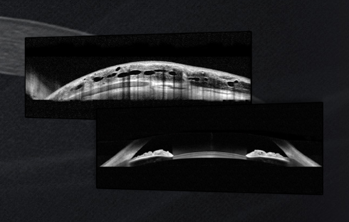
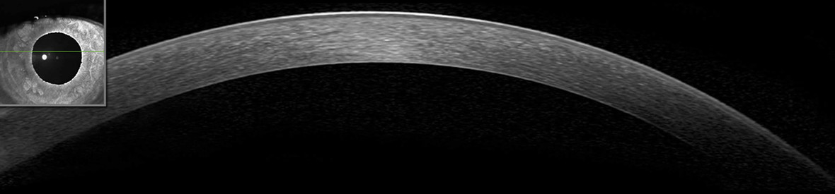
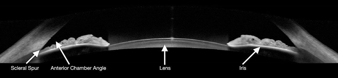
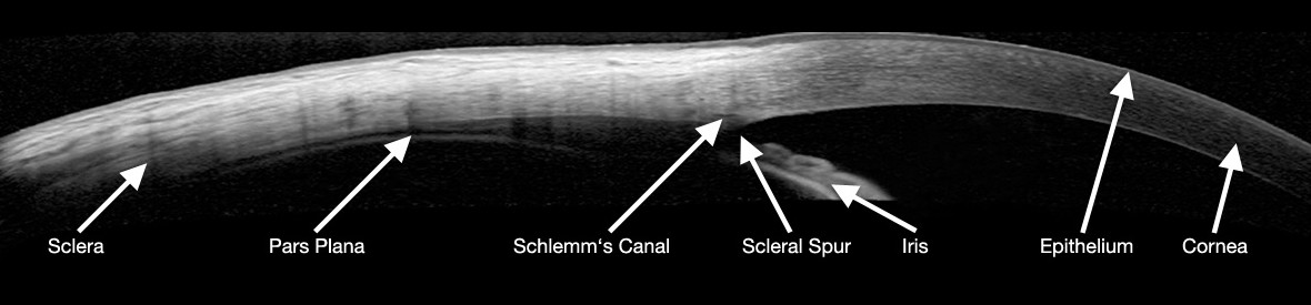
ANTERIOR SEGMENT MODULE
High-resolution anterior segment imaging
The high-resolution images obtained with the SPECTRALIS¢î Anterior Segment Module show the layers of the cornea in detail and aid in the assessment of corneal thickness.
The module also offers angle-to-angle imaging for efficient anterior chamber angle evaluation as well as detailed images of scleral structures.
Glaucoma Module Premium Edition
The Spectralis Glaucoma Module Premium Edition combines the proprietary Anatomic Positioning System (APS) with a series of unique scan patterns to assess the optic nerve head, the retinal nerve fiber layer, and the macular ganglion cell layer.
These APS-based scan patterns are automatically matched to each eye¡Çs unique anatomic landmarks and to the characteristics of fine anatomic structures relevant in glaucoma diagnostics.
The Glaucoma Module Premium Edition compares patients¡Ç eyes to a reference database of normal eyes, noting even very small deviations. The precision of the SPECTRALIS AutoRescan function allows confident identification and monitoring of structural changes from visit to visit.
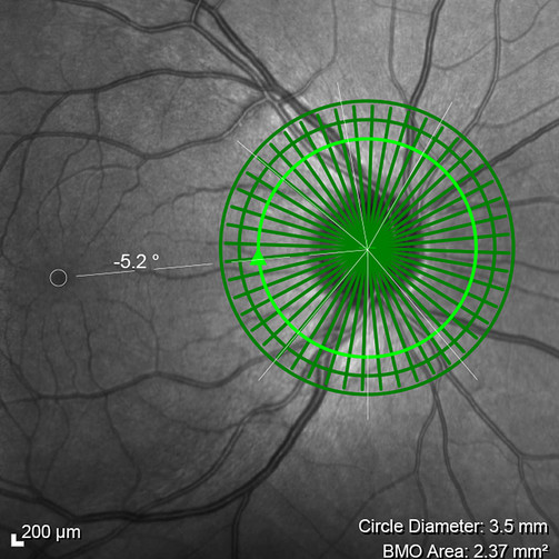
Anatomic Positioning System
SThe patented Anatomic Positioning System (APS) creates an anatomic map of each patient¡Çs eye using two fixed, structural landmarks: the center of the fovea and the center of Bruch¡Çs membrane opening. With APS, all scan protocols are automatically oriented according to the patient¡Çs anatomic map.
This enables precise examination of relevant structures and ensures accurate comparisons with reference data, allowing for a highly sensitive assessment of structural change.
Lorem Ipsum
Lorem Ipsum is simply dummy text of the printing and typesetting industry. Lorem Ipsum has been the industry's standard dummy text ever since the 1500s, when an unknown printer took a galley of type and scrambled it to make a type specimen book. It has survived not only five centuries, but also the leap into electronic typesetting, remaining essentially unchanged. It was popularised in the 1960s with the release of Letraset sheets containing Lorem Ipsum passages, and more recently with desktop publishing software like Aldus PageMaker including versions of Lorem Ipsum.